A BOUTIQUE MAMMOGRAPHY EXPERIENCE
Camellia Women’s Imaging provides the women of Alabama the best and latest technology for breast imaging and bone density in a comfortable setting. Our breast imaging technology has been selected to maximize our success in the detection of breast cancer and breast disease, while minimizing your anxiety. We want to make your visit with us a pleasant memory. You may contact us online or call us (205-544-2828).
Screening
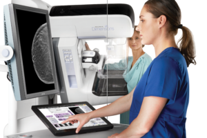
A screening mammogram is a special x-ray picture of the breast performed in women without symptoms to allow for the early diagnosis of breast cancer or other breast abnormalities. This can be done with or without 3D imaging. The high-quality mammogram technology used at Camellia is provided with delicate care, and above all, kindness to our patients. Screening mammograms are usually performed in women who do not have any symptoms or signs of breast cancer. Higher risk women or women who have previously had breast cancer can also get screening mammograms if they are not having any symptoms. At Camellia, we provide mammograms with state-of-the-art technology in a beautiful, calming, and friendly environment where stress is minimized.
Currently yearly mammograms are recommended beginning at age 40 for all women. Earlier yearly screening may be recommended in women who are at a higher risk for breast cancer or have a family history of breast cancer. Please call us to determine which type of mammogram is best for you based on your symptoms, risk, or history.
The radiologist will read your mammogram while you are waiting and discuss the results of your screening with you the day of your exam. Also, if there is a need for further views or ultrasound, these can usually be performed on the same day. We strongly believe that quality time should be spent with all patients.
To request an appointment, please click here.
Diagnostic
Diagnostic mammograms are performed when you have a symptom such as a lump felt by you or your doctor, thickening in your breast, nipple discharge, change in breast size or shape, or localized pain in the breast. Diagnostic mammograms can also be performed in some cases if you have had a abnormal screening mammogram, a personal breast cancer history, or as a follow-up to an abnormal mammogram.
We use the latest Hologic conventional digital and 3D mammography and if needed, high resolution ultrasound to perform diagnostic breast imaging. When you come in for a diagnostic mammogram, the radiologist will read your mammogram while you are waiting, and specialized or additional views will be performed the same day, if needed. The radiologist will talk with you before you leave to explain your study results, answer any questions, and help you coordinate any follow-up. If you need any additional appointments we will help you make them.
To request an appointment, please click here.
Camellia offers 3D mammography technology or breast tomosynthesis. This is a revolutionary technology that can be used for screening or diagnostic mammograms. In a traditional 2D mammogram the radiologist looks at an image where all of the breast tissue is superimposed. With 3D mammography the breast is imaged in thin slices. The images can be looked at like pages in a book. Studies have shown that 3D mammography allows for higher cancer detection rates and lower false positive rates. The procedure to get 3D will feel the same as standard mammography. Additionally, with 3D mammograms there is often a decreased need for additional mammogram views or “callbacks” for benign or incidental findings.
The radiation in a 3D mammogram is slightly greater than with 2D but it is well within the allowable safe standards of the FDA and ACR. Additionally, Camellia is one of the few centers to offer C-view, a technology that can reduce the radiation does. Based on recent evidence, it seems that the 3D performs better than 2D mammograms for cancer detection. We chose these technologies because we want you to feel so cared for that you tell your best friend about us.
For additional information on 3D Mammography, please click here.
To request an appointment, please click here.
Camellia’s Hologic mammogram machine provides digital imaging. Digital mammography has been around since approximately 2005. It allows radiologist to read your mammogram on a very high-resolution computer screen rather than on film. Digital mammograms can be 2D mammograms, 3D mammograms, or a combination of both. Digital mammography machines are designed to tailor the amount radiation for each person, so that the best images are obtained using the minimum amount of radiation needed. Because the images are digital, and read on a computer, this allows for the use of a technology called Computer Aided Detection (CAD). CAD draws the radiologist’s attention to areas on the mammogram that may contain suspicious calcifications or masses. We take the time to provide and review this technology because we want to treat you the way we would treat our family.
To request an appointment, please click here.
Screening
Breast ultrasound is used in conjunction with mammography to evaluate the breast. Camellia Imaging offers a high resolution Siemens S2000 ultrasound machine. Breast ultrasound does not require the breast to be compressed or require any radiation exposure. Breast ultrasound is not typically used for screening in all women. In some cases, however, it may be beneficial for screening, especially in women with dense breast tissue. We offer screening breast ultrasound to women without symptoms with denser breast tissue and some risk factors for breast cancer. We always tailor our services to meet your individual needs. We will discuss all results with you at your appointment, answer any questions you may have, and communicate those results to your doctor.
To request an appointment, please click here.
Diagnostic
Sometimes ultrasound is used as a diagnostic problem-solving tool for the breast and underarm area. It can be used to evaluate something that is felt by you or your doctor, or something seen on you mammogram. The ultrasound examination done in conjunction with the mammogram and/or clinical exam can help better characterize the area of interest. It can sometimes also help evaluate for rupture of breast implants. We offer high resolution Siemens S2000 ultrasound performed by our technologist and radiologist. We always tailor our ultrasound services to meet your individual needs. We will discuss all results with you at your appointment, answer any questions you may have, and communicate those results to your doctor.
To request an appointment, please click here.
Needle Biopsy
We perform needle biopsies of the breast in the comfort of our office, using our state-of –the –art imaging tools. Biopsies can be preformed in a minimally invasive manner using ultrasound, stereotactic or 3D (tomosynthesis) guidance. We perform needle biopsies with and without vacuum assistance. We also perform cyst aspirations and fine needle aspirations. We will walk you through the procedure step by step to ease your anxiety. We want you to feel comfortable with your procedure. Our equipment allows us to see and accurately target the area that requires biopsy. We usually place a tiny biopsy clip at the time of biopsy to mark the spot of the biopsy. The biopsy samples are sent to a trusted breast pathologist, and we will explain your results to you and answer your questions within one or two business days after the biopsy.
Ductogram / Galactogram
For women who have nipple discharge or secretion that is suspicious, we perform a ductogram or galactogram to determine if something is seen in the duct that could explain this abnormal discharge. A small catheter can be introduced in to the duct. Injection of contrast into this small catheter can help us see the contour of the duct. If an abnormality is seen on the ductogram, we can use our mammography biopsy equipment to help biopsy and determine the cause of the discharge in a minimally invasive manner.
Needle Localization
If there is something found by biopsy in the breast that requires removal by a surgeon because it is breast cancer, pre-caner, high risk, or indeterminate we can perform a needle localization or a wire placement. Before the surgery, a wire or multiple wires are inserted into the breast to help guide the surgeon the area or areas to be surgically removed. This allows the surgeon to be more precise and remove the area of concern. This procedure can be performed with mammographic or ultrasound guidance. The procedure can be performed in the comfort of our office the day of the surgery. We recognize that breast surgery can be very stressful. We do everything we can to help alleviate as much of this stress as possible.
Bone density is an important part of caring for a women’s health because when women age, have removal of their ovaries, or take certain medication their bone density can decrease. Very low bone density is called osteoporosis, and can increase your risk of fractures. Because fractures can be debilitating, measurement of bone density to allow for treatment of osteoporosis is very important to prevent unnecessary fractures. Camellia Women’s Imaging provides bone density evaluation with the most advanced Hologic A Series DEXA Unit. DEXA can evaluate for bone density to diagnose osteoporosis and can evaluate for fractures in the mid and lower spine. The results are explained at the time of the examination by the doctor. These results will also be sent to your doctor so they can guide your therapy. Please do not take your calcium supplement for at least one day prior to your bone density evaluation.
To request an appointment, please click here.
Camellia Women’s Imaging is one the few places in Alabama to offer the latest in DEXA body composition scanning, a fast, highly accurate test to measure muscle, fat, and bone content. DEXA is considered the gold standard for body composition analysis. Body Composition Analysis (BCA) details where fat and lean muscle are distributed throughout the body and provides a comprehensive color report summarizing the study which will be explained to you by a doctor on the day of the examination. Additionally, it can also measure the about of visceral adipose tissue or fat surrounding the organs which can indicate ones risk for cardiovascular disease and diabetes. It is often a helpful tool for individuals starting anew diet or workout regimen.
To request an appointment, please click here.